An adorable baby

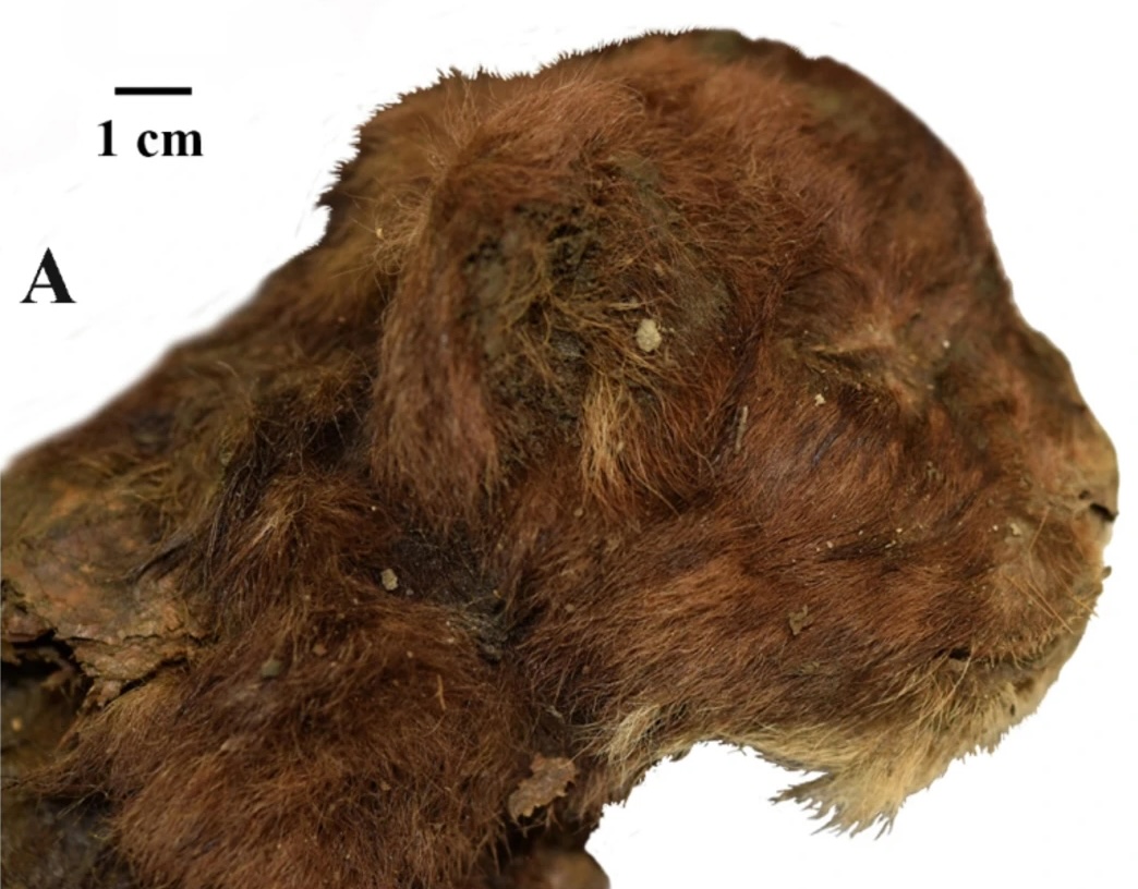
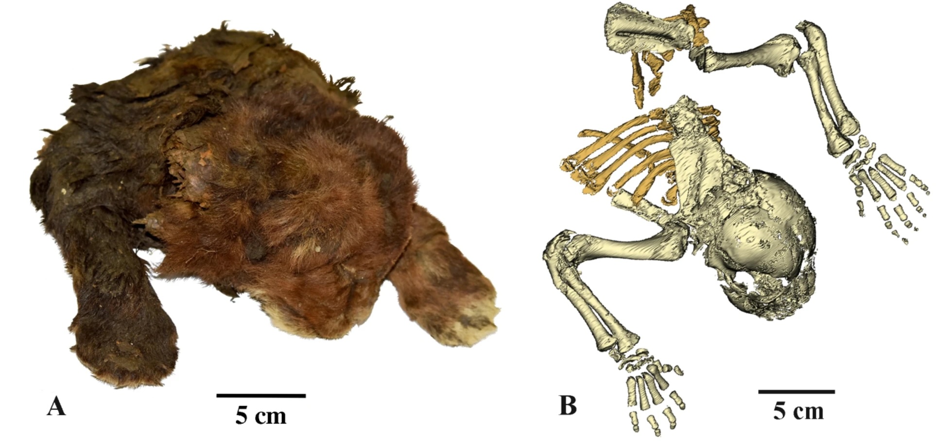
What a cute little dead baby.

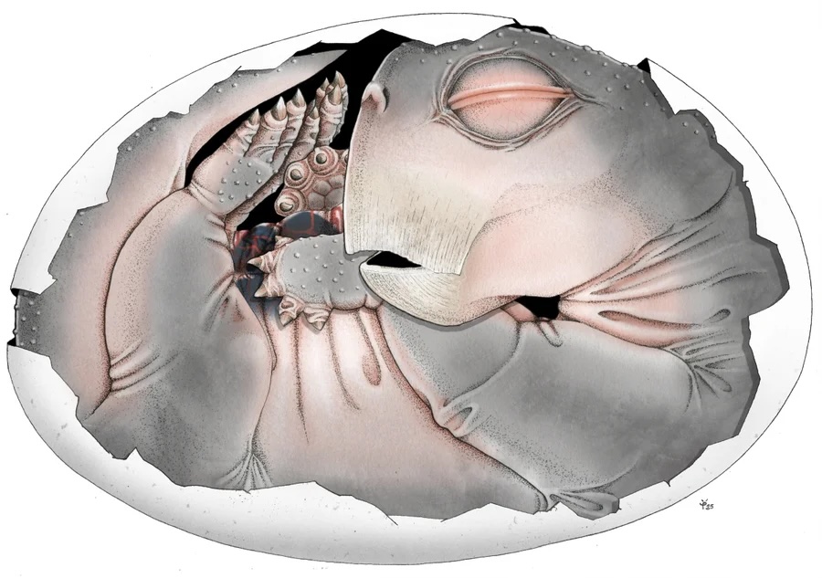
That’s a Lystrosaurus embryo. If you don’t know Lystrosaurus, it’s an amazing species that survived the Permian extinction and experienced a remarkable population boom — it was a large vertebrate that came to dominate the planet after that mass extinction, yet most people have never heard of them. We ought to pay more attention! Memento mori, and all that.

Anyway, this fossil answers the least surprising question ever.

Detailed imaging of a 250-million-year-old fossil has revealed the first proof that the ancestors of mammals laid eggs. The discovery answers a long-standing question about the reproductive biology of our ancient forerunners and hints at how they managed to flourish in the aftermath of the biggest mass extinction in Earth’s history.

Scientists have long assumed that the ancestors of mammals—a group known as the therapsids—laid eggs like today’s platypuses and echidnas do. But they lacked any direct evidence of synapsid eggs in the fossil record.

It’s good to be able to tick off that one box, confirming that Permian therapsids laid eggs, but it’s hardly news. I thought this was much more interesting:

Most importantly, the new images reveal that the two halves of the lower jaw had yet to fuse in the youngest Lystrosaurus specimen. In turtles and birds, the lower jaw fuses before birth, allowing the baby to feed itself after hatching. The unfused lower jaw of this Lystrosaurus is therefore another indication that the animal died while still in its egg. The other two specimens exhibit signs of having been somewhat more mature; the largest one was preserved in a splayed-out posture that shows it was not in an egg and had traveled some distance before dying.

Mammalian ancestors invested more in maternal care than some other organisms. That might be a clue to how our clade survived through a couple of mass extinctions.

I’m not the only one easily seduced by giant Cretaceous octopuses

You knew I’d have to read an article titled Earliest octopuses were giant top predators in Cretaceous oceans. How cool is that? And then they’ve illustrated it with some very appealing figures.

Body size estimation of Late Cretaceous octopuses.
The graph shows an allometric relationship between the length of the jaw and mantle in long-bodied species of extant finned octopus . The name of the corresponding species is shown along each growth curve. The sizes of N. jeletzkyi and N. haggarti based on their largest specimen are indicated by black vertical lines. Reconstruction of these two species, the extant giant squid, and gigantic vertebrate predators in the Late Cretaceous are shown with their maximum total length.

Also, the abstract promises much.

Top predators drive changes in ecosystem structure. For the last ~370 million years, large-sized vertebrates have dominated the apex of the marine food chain, while invertebrates have served as smaller prey. Here we describe invertebrate top predators from this “age of vertebrates,” the earliest finned octopuses (Cirrata) from Late Cretaceous sediments (~100 to 72 million years ago), as identified based on huge, exceptionally well-preserved fossil jaws and their wear. This extensive wear suggests dynamic crushing of hard skeletons. Asymmetric wear patterns further indicate lateralized behavior, suggesting advanced intelligence. With a calculated total length of ~7 to 19 meters, these octopuses may represent the largest invertebrates thus described, rivaling contemporaneous giant marine reptiles. Our findings show that powerful jaws, and the loss of superficial skeletons, convergently transformed cephalopods and marine vertebrates into huge, intelligent predators.

But does the paper deliver? Sad to say, it doesn’t. I was disappointed on how far the authors stretched an interesting technique to reach an excessive conclusion.

What they did was collect fossil octopus beaks and subject them to grinding tomography — basically shaving away the rock, photographing each exposed slice, and using an AI to help reconstruct a detailed 3-D image of the beak that allowed them to view the wear and tear on the beak’s surface, presumably seeing the damage acquired as they chewed their way through their Cretaceous prey. The entirety of the data in the paper is an analysis of scratches and wear on these beaks.

Huge lower jaws of fossil octopuses and of an extant giant squid.
(A and B) The largest lower jaws of the Late Cretaceous finned octopus species N. jeletzkyi [(A) NMNS DS00042 3LmvTpM] and N. haggarti [(B) KMNH IvP 902001]. Both specimens show extensive loss of jaw material caused by wear. (C) A lower jaw of the extant giant squid Architeuthis dux (NSMT-Mo 85956), a species having the largest jaw among modern cephalopods. (A) is a digital fossil jaw visualized as a 3D model; (B) is an exceptionally well-preserved nondigital fossil jaw; and (C) is a modern jaw dissected from a carcass of ~10 m total body length. Solid lines indicate the extension of striation on the outer surface of the hood and broken lines show the estimated outline of the rostrum without wear. The hood and lateral walls lost by weathering, shown as shadowed areas, are reconstructed based on the holotype and specimens in fig. S4. (A) and (C) are exhibited in a mirrored position. Scale bar, 20 mm.

That’s good stuff. No data too small — it’s all data. But wait: this paper contains nothing but measurements of beaks, but manages to expand this into a whole set of conclusions about the marine ecosystem.

These wear patterns suggest that Late Cretaceous giant Cirrata were active carnivores that frequently crushed hard shells and bones. The long scratches distributed on wide areas of their jaw reflect the dynamic use of the entire jaw for dismantling prey. Asymmetric loss of the jaw edges suggests lateralized behavior, which has been linked to a highly developed brain and cognition. This, in turn, suggests that the earliest octopuses already possessed advanced intelligence. Laterality is known in modern octopuses, whose high intelligence matches that of vertebrates. The exceptionally large jaws of adult N. jeletzkyi and N. haggarti suggest a strong bite force because cephalopod jaw muscles enlarge as the jaw size increases. The long lateral walls in their jaws revealed by the new digital specimens reported here show that Nanaimoteuthis had large jaw muscles. The chipping on both the rostrum and jaw edge was caused by strong shear stress beyond the yield point of the most robust part of the jaw. The transverse cracks in N. haggarti are probably a trace of larger shear failures. These large fractures thus suggest a powerful bite. In giant Cirrata, the jaws are smaller than those of contemporaneous Cretaceous vertebrate top predators, which measure ~1.7 m in length. Instead of using a large mouth, the long and flexible arms of octopuses serve for catching large prey. The giant Cirrata probably consumed large prey with their long arms and jaws, playing the role of top predators in Cretaceous marine ecosystems.

All we’ve got are scratches on beaks, with extrapolation from beak size to overall size. From that we leap to the conclusion that these giant octopuses were rivals to mosasaurs, plesiosaurs, ichthyosaurs, and sharks. We assume they’re top predators in the absence of actual evidence of predation or their role in the ancient ecosystem.

Convergent evolution among marine top predators in the Paleozoic–Mesozoic.
This model shows the acquisition of jaws and the reduction of superficial skeletons in the evolutionary history of marine vertebrates (top) and cephalopods (bottom) to become top predators. The gray horizontal bars show the chronological range of some selected groups of vertebrates and cephalopods. For cephalopods, stepwise reductions of skeletons are indicated by the blue background.

I’m not even going to touch the idea that asymmetric scratches are good evidence of high intelligence.

I am reminded of the speculations of Mark McMenamin, who thought circular shapes in Triassic sediments were evidence of a gigantic Kraken. He also found a broken piece of rock that he extrapolated to claim it was the tip of a giant kraken beak.

At least McMenamin’s extravagant conclusions weren’t getting published in Science.

Mother of spiders!

Show me a 500 million year old chelicerate, and I’ll be happy for a day. Look at this beauty, Megachelicerax cousteaui, excavated from a Utah fossil bed.

Anatomic reconstructions of the dorsal (left) and ventral (right) morphologies. b, Artistic reconstructions by M. Hattori illustrating oblique views of the dorsal (top) and ventral (bottom) morphologies. The sanctacaridid-like morphology of the posteriormost body region is speculative. gi, gill (that is, a set of gill lamellae); te, telson.

Pretty cool, right? The best part of it is that pair of appendages at the very front of the animal — those are chelicerae, the biting/chomping/chewing/venom-injecting bits of a modern spider, that make them distinct from insects, which only have antennae at that end. That makes this the oldest known chelicerate ever discovered. It was a swimming marine animal, and doesn’t have the legs we associate with spiders — chelicerae evolved first, legs much later.

Also, this isn’t just the mother of spiders, but is also the mother of a huge family of cousins: horseshoe crabs, eurypterids, as well as spiders.

Megachelicerax documents the oldest stratigraphic occurrence of chelicerae (that is, uniramous, unichelate deutocerebral appendages) and bridges the simple body and limb organization of Cambrian megacheirans with the more derived anatomy of post-Cambrian synziphosurines and crown-group chelicerates. a, Simplified consensus topology based on Bayesian analysis (Mk model, 4 chains, 5,000,000 generations, 1/1,000 sampling resulting in 5,000 samples with 25% burn-in resulting in 3,750 samples retained); detailed results and comparison with parsimony provided in Extended Data Fig. 6. The numbers in parentheses correspond to the total number of podomeres and the number of chelae, respectively, present in the deutocerebral appendage. Taxa whose names are in bold font are illustrated in b–l. b–l, The morphology of the anterior body region in select taxa. b, Fuxianhuiid Chengjiangocaris kunmingensis (Cambrian, Stage 3). c, Artiopod Olenoides serratus (Cambrian, Wuliuan). d, Megacheiran Yohoia tenuis (Cambrian, Wuliuan). e, Megacheiran Haikoucaris ercaensis (Cambrian, Stage 3). f, Megacheiran Leanchoilia superlata (Cambrian, Wuliuan). g, Mollisoniid M. plenovenatrix (Cambrian, Wuliuan). h, Habeliid Habelia optata (Cambrian, Wuliuan). i, M. cousteaui (Cambrian, Drumian). j, Synziphosurine Dibasterium durgae (Silurian, Wenlock). k, Xiphosurid Limulus polyphemus (recent). l, Eurypterid Slimonia acuminata (Silurian, Llandovery–Wenlock).

That is one wildly successful tree. It just goes to show that you can go on to do great things even if your face looks like a nest of spiky clawed jointed tentacles.


Rudy Lerosey-Aubril, Javier Ortega-Hernández. A chelicera-bearing arthropod reveals the Cambrian origin of chelicerates. Nature, 2026; DOI: 10.1038/s41586-026-10284-2

Wanna see a beautiful fossil?

I know that a pile of bones of Tyrannosaurus or Triceratops gets all the attention and popular press, but what gives me a thrill is seeing a well-preserved Cambrian invertebrate, especially if it represents an early developmental stage. Here’s a real beauty, the phosphatized larva of Youti yuanshi, from Yunnan, China.

YKLP 12387. a, External scanning electron microscopy, right side. Damage to posterior epidermis exposes lining of perivisceral cavity, demonstrating blind gut. b, External scanning electron microscopy, left side. c,g–j, Median virtual dissection from X-ray computed tomography (XCT) data (c), showing location of transverse slices intersecting digestive glands (g,i) and transverse membrane (h,j). d, Semi-manual segmentation of internal chambers from XCT data, viewed from the left side. Dorsolateral aspects of the peripheral cavity are omitted for clarity. e,f, Virtual dissection parallel to coronal plane, looking ventrally (e) and dorsally (f), showing digestive glands, pericardial sinus, transverse membranes within perivisceral cavity, and oblique membranes within peripheral cavity. g–j, XCT sections at positions indicated in c at position of digestive glands (g,i) and at position of ventrolateral lacunae and transverse membrane (h,j). g,h, Sections close to the anterior trunk, reflecting segments at late developmental stage. i,j, Sections close to the posterior trunk, showing superior preservation of internal tissue. k, Segmentation of internal chambers from XCT data, viewed from the dorsal perspective at anterior, middle and posterior trunk. Aspects of peripheral cavity are omitted for clarity. a, appendage; cb, central body of brain; db, dorsolateral body of brain; dia, diagenetic grain; dg, digestive gland; dm, dorsal membrane; dp, dorsal projection; dv, dorsal vessel; fb, frontal body of brain; irr, irregular chamber; lig, ligament; om, oblique membrane; pc, pericardial sinus; pph, peripheral cavity; pn, perineural sinus; pv, perivisceral cavity; tm, transverse membrane; vl, ventrolateral sinus; vv, ventral vessel. Scale bars, 200 μm.

Superficially, it looks like a grub you might dig up in your garden, but this was found in marine sediments and was less than 4mm long, so you’d be unlikely to find anything like it today. It’s from a paper titled Organ systems of a Cambrian euarthropod larva by Martin R. Smith, Emma J. Long, Alavya Dhungana, Katherine J. Dobson, Jie Yang & Xiguang Zhang. The specimen is so well preserved that it can be studied at the level of organs and organ systems.

The Cambrian radiation of euarthropods can be attributed to an adaptable body plan. Sophisticated brains and specialized feeding appendages, which are elaborations of serially repeated organ systems and jointed appendages, underpin the dominance of Euarthropoda in a broad suite of ecological settings. The origin of the euarthropod body plan from a grade of vermiform taxa with hydrostatic lobopodous appendages (‘lobopodian worms’) is founded on data from Burgess Shale-type fossils. However, the compaction associated with such preservation obscures internal anatomy. Phosphatized microfossils provide a complementary three-dimensional perspective on early crown group euarthropods, but few lobopodians. Here we describe the internal and external anatomy of a three-dimensionally preserved euarthropod larva with lobopods, midgut glands and a sophisticated head. The architecture of the nervous system informs the early configuration of the euarthropod brain and its associated appendages and sensory organs, clarifying homologies across Panarthropoda. The deep evolutionary position of Youti yuanshi gen. et sp. nov. informs the sequence of character acquisition during arthropod evolution, demonstrating a deep origin of sophisticated haemolymph circulatory systems, and illuminating the internal anatomical changes that propelled the rise and diversification of this enduringly successful group.

Here’s a helpful diagram to help sort out what’s going on inside the worm.

a, Organ system disposition in sagittal view. Dotted lines denote location of sections shown in e,f. b, Organ system disposition in transverse view. c,d, Head, from lateral perspective (c) and as medial transverse section (d). e,f, Transverse sections through trunk at location of digestive glands (e) and transverse membranes (f). g–j, Coronal sections through head, from ventral (g) to dorsal (j) planes. Colour scheme as in Fig. 1.

Most interesting is the comparative analysis with other Cambrian organisms, especially with regards to the organization of the nervous system.

Phylogenetic analysis situates Youti yuanshi within the AOPK clade containing Anomalocaris, Opabinia, Pambdelurion and Kerygmachela. Under our preferred model, the circumoral brain ring of cycloneuralians corresponds to the panarthropod prosocerebrum, which innervates the first appendage pair (onychophoran antennae, tardigrade stylets or euarthropod labrum). We interpret the archicerebrum as a distinct development dorsal to the prosocerebrum, associated with sensory receptors: specifically the eyes, and the dorsal projections (Kerygmachela rostral spines, tardigrade cirri, crustacean frontal filaments or anterior paired projections of stem euarthropods; homology with the anteriormost onychophoran lip papillae is plausible, but may not be parsimonious). The taxa depicted in this figure are selected in order to depict the evolutionary context of Youti; the relationships shown are recovered under all analytical conditions.

Over half a billion years ago, the oceans were filled with diverse wormlike animals that were exploring different arrangements of their squishy bits — it was a complex ecology and every individual discovered should fill us with awe. Each of these forms has a deeper history that we need more fossils to decipher.

Not the prettiest jumping spider I’ve seen

I’m used to seeing spectacularly pretty Australasian jumping spiders, and this one, the genus Simaetha, isn’t exactly dazzling.

Australian representatives of the two extant Simaethina genera: A, C, E, Simaetha sp. (female); B, D, F, Simaethula sp. (female). Specimens are shown in dorsal view (A, B), lateral view (C, D) and frontal view (E, F). Scale bars: 0.5 mm (B, D, F), 1 mm (A, C, E).

This one, though, has the excuse that it’s between 11 and 16 million years old. It isn’t that old — I’d expect that the planet had lot of jumping spiders during the miocene — but it’s nice to seen an example from that period.

Simaetha sp. indet. (AM F.161027). Only known specimen: A, light microphotograph. B, scanning electron micrograph. C, morphological interpretation of light and electron micrographs. Abbreviations: LL, left leg; RR, right leg; AME, anterior median eye; PME, posterior median eye. Scale bars: 0.5 mm.

It’s also impressive that they could sort out what was what in the squashed bits of that fossil.

BIRDIE!

Time to dig up another fossil. It’s a bird, but it has no connection to the sabre-toothed kitty cat I posted yesterday — this is a 80 million year old bird, Navaornis hestiae, written up in a Nature article, Cretaceous bird from Brazil informs the evolution of the avian skull and brain. It looks like a real bird to me.

a,b, Photograph (a) and interpretive drawing (b) of the exposed side of the holotype of N. hestiae (MPM-200-1) in left lateral view. c, Micro-computed tomography rendering of MPM-200-1 in right ventral–lateral view. Scale bar, 10 mm.

It has a fairly big brain, with some differences in structure from modern birds — it has a smaller motor control area, so while it had the capacity for complex behavior, it may not have been as agile in the air as birds today. It’s intermediate in brain complexity between Archaeopteryx and extant birds.

a, Three-dimensional reconstruction of the endocranial morphology of N. hestiae from MPM-200-1 and MPM-334-1. Portions deriving from MPM-200-1 and MPM-334-1, as well as the reconstruction process, are explained in the Methods and Extended Data Fig. 7. b, Evolution of endocranial morphology across Pennaraptora. Numbers in the coloured boxes refer to the degree of expansion of each of the main neuroanatomical and sensorial regions for each taxon. Brown arrows in b depict the orientation of the foramen magnum.

Cambridge invested a bit in publicizing this discovery, with a nice fancy video.

KITTY!

External appearance of three-week-old heads of large felid cubs, right lateral view: (A) Homotherium latidens (Owen, 1846), specimen DMF AS RS, no. Met-20-1, frozen mummy, Russia, Republic of Sakha (Yakutia), Indigirka River basin, Badyarikha River; Upper Pleistocene

I understand that the internet likes cats, so here’s one, a 30,000 year old mummified sabre-toothed kitten.

It has a distinctively large mouth and massive neck muscles, but the canine teeth haven’t elongated yet — they say the age is equivalent to a 3 week old lion cub. I would guess that sabre-tooth canines might interfere with nursing.

The frozen mummy of Homotherium latidens (Owen, 1846), specimen DMF AS RS, no. Met-20-1, Russia, Republic of Sakha (Yakutia), Indigirka River basin, Badyarikha River; Upper Pleistocene: (A) external appearance; (B) skeleton, CT-scan, dorsal view.

It has toe-beans!

Bring me the head of Arthropleura

We’ve known about these amazing fossils from the lower Carboniferous for a while — it’s Arthropleura, a gigantic 2.5 meter long millipede. Imagine cleaning up your kitchen when a beast 2 or 3 times your length fluidly, sinuously crawls out from your baseboards. Wouldn’t that be neat?

One of the only problems with imagining that is that none of the fossils to date have had a head. Sure, it’s imposingly large, but what kind of face does it have? It’s a millipede, and millipedes are harmless detritivores who aren’t going to be a threat at all, unless you’re a pile of moldering leaves or a fungus. It’s centipedes that are primarily carnivores, with pointy sharp venomous forcipules that can deliver a nasty bite. That Arthropleura is in the millipede clade tends to blunt their potential menace.

Good news, time-traveling super-villains looking for a pet! The head of Arthropleura has at last been discovered, and it’s centipede-like, with strong bitey jaws, and also has stalked eyes. It’s a bit squished.

(A and B) Three-dimensional reconstruction. (A) Dorsal view. (B) Ventral view. (C and D) specimen inside the nodule. (C) Part. (D) Counterpart. Co, collum; DT, digestive tube; H, head; Pt, paratergite; S#, sternite number; St, syntergite; T#, tergite number; Te, telson. Reconstructions are made from Phoenix X-ray Phoenix V|tome|x CT scan. Scale bars, 1 cm (C and D) and 5 mm (A and B).

(A) Dorsal view. (B) Ventral view. (C) Back view. (D) Frontal view. Left maxillae were removed on (B) to better illustrate the mandible below. The red circle on (C) indicates the position of the digestive tract.

However, it’s still thought likely that it was a detritivorous. This has advantages for those of us who really want one as a pet: it’s still an intimidating creature, but in its free time it can roam the lair, cleaning up any untidiness.

Yes, I might fantasize a bit about keeping a few Arthropleura about the house. Better than a dog, anyhow.

These monsters are all dead

I hope you all like long tubular creatures, because that’s all I’ve got for you today. Maybe they’d be less horrifying if they had lots of legs?

Here’s a 4-meter long salamander-like beast from the Permian, named Gaiasia.

I’ve seen giant salamanders before, but not ones with big box-like skulls full of razor-sharp fangs.

Here’s another muscular tube, Vasuki indicus, only 47 million years old, but somewhere around 10-15 meters long.

The amusing thing about this beast is that everyone in the popular press treatment is making it all about how long it is — it’s a partial skeleton, there’s not enough to determine exactly how long it is. It’s either shorter than Titanoboa, the gold standard of giant ancient snakes, or bigger than Titanoboa. It’s not a competition, people! They’re separated by about 10 million years. But of course they’re in competition for starring roles in cheesy sci-fi CGI epics.

That’s why we’re seeing ridiculous comparisons like this one:

OK, the snake was longer than T. rex, but so what? It wasn’t as massive, and they were temporally distant from one another. This illustration reveals how some people are thinking:

That could be an ad for the next movie by The Asylum. These kinds of team-ups are popular to promote cheese, like Godzilla vs. Mechagodzilla or Dracula vs. Frankenstein. Learn to love Vasuki for itself, OK?

A Carboniferous arachnid

This week has been a good one for chelicerate evolution. Here’s another fossil, Douglassarachne acanthopoda, which was creeping around in the forests of Illinois in the late Carboniferous.

Douglassarachne acanthopoda n. gen. n. sp., holotype and only known specimen FMNH PE 91366; for interpretative drawings and scale, see Figure 2. (1) Part, detail of distal femur and more-distal podomeres, showing nature of curved macrospines on lateral edge of distal podomeres, bases of macrospines on dorsal surface of femur; (2) counterpart, detail of posterior opisthosoma showing bilobed structure at base of anal tubercle.

What is it? I don’t know. The authors are unsure. It’s an arachnid, but it could be in the spider lineage or the harvestman lineage, or it could be its own weird thing. It’s spiderish, anyway.

Douglassarachne acanthopoda n. gen. n. sp., reconstruction of the possible appearance of the animal in life.