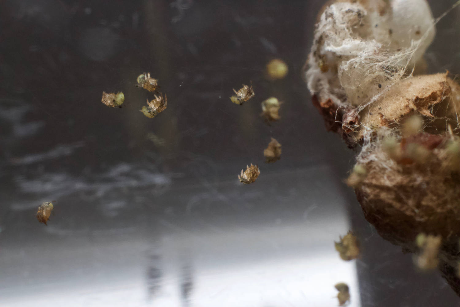
My colleague, Chris Atkinson, told me yesterday that he’d been seeing a lot of spiders in his compost heap. “Interesting,” I thought. Then he sent me this photo:
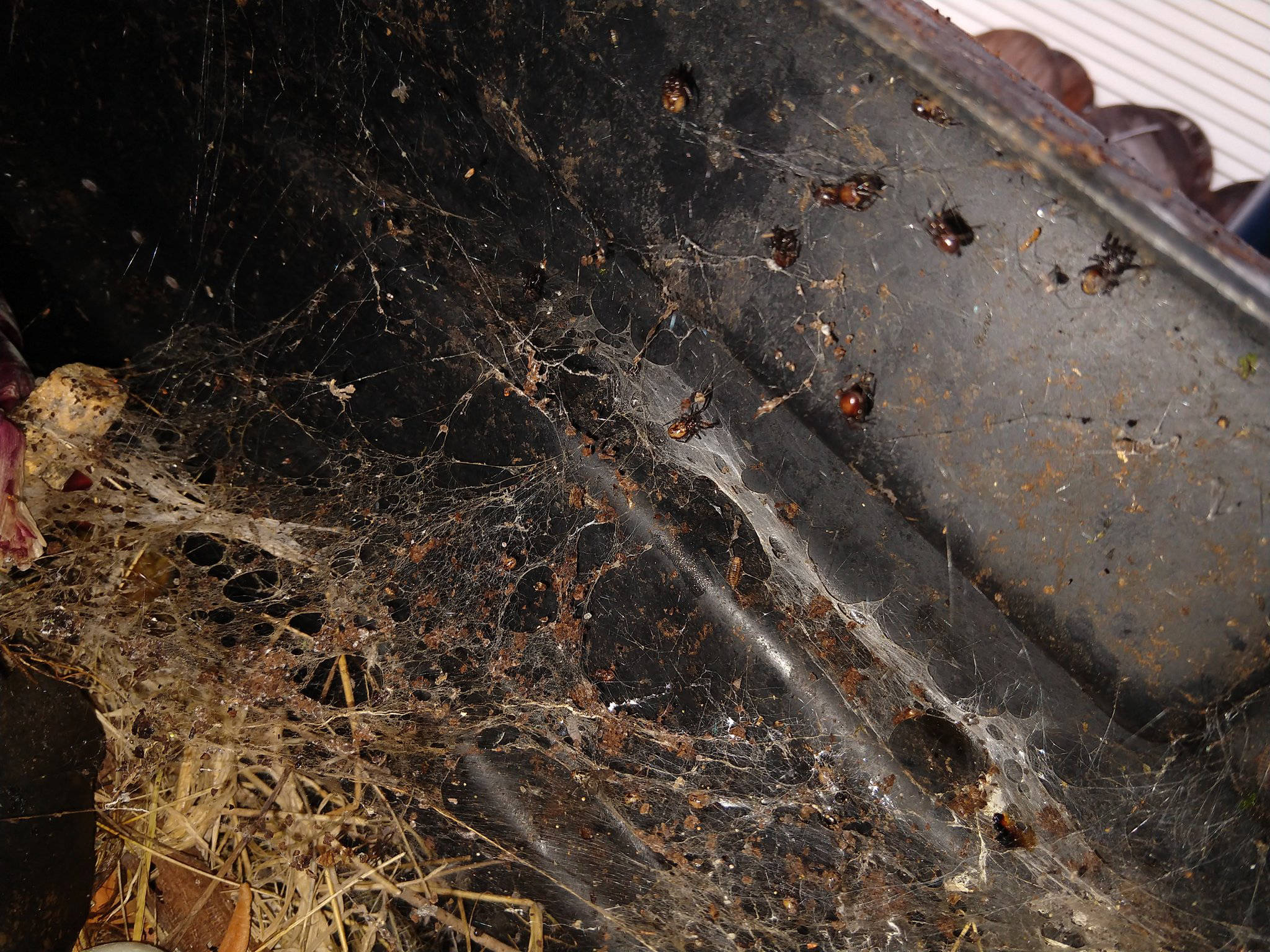
WHOA. Look at all those spiders.
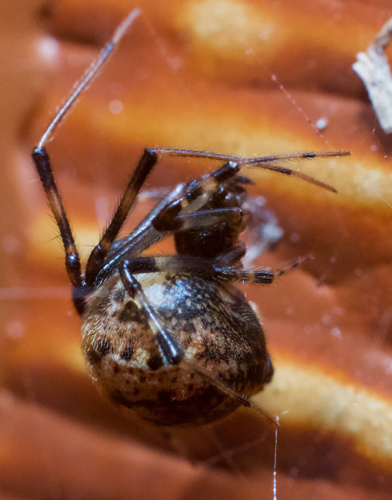
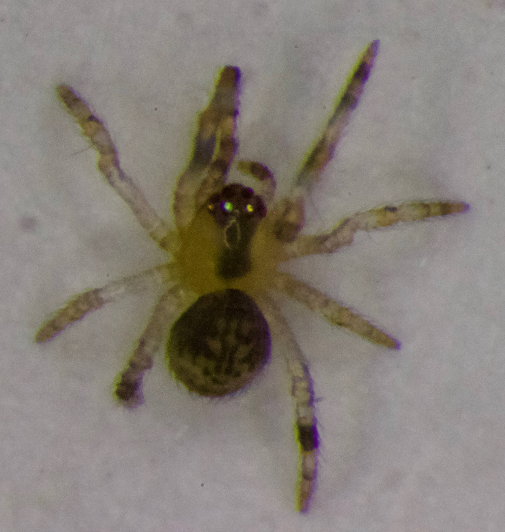
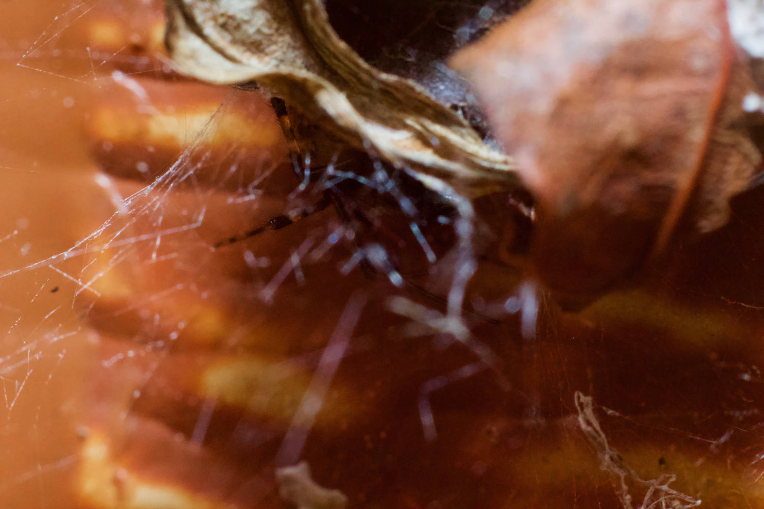
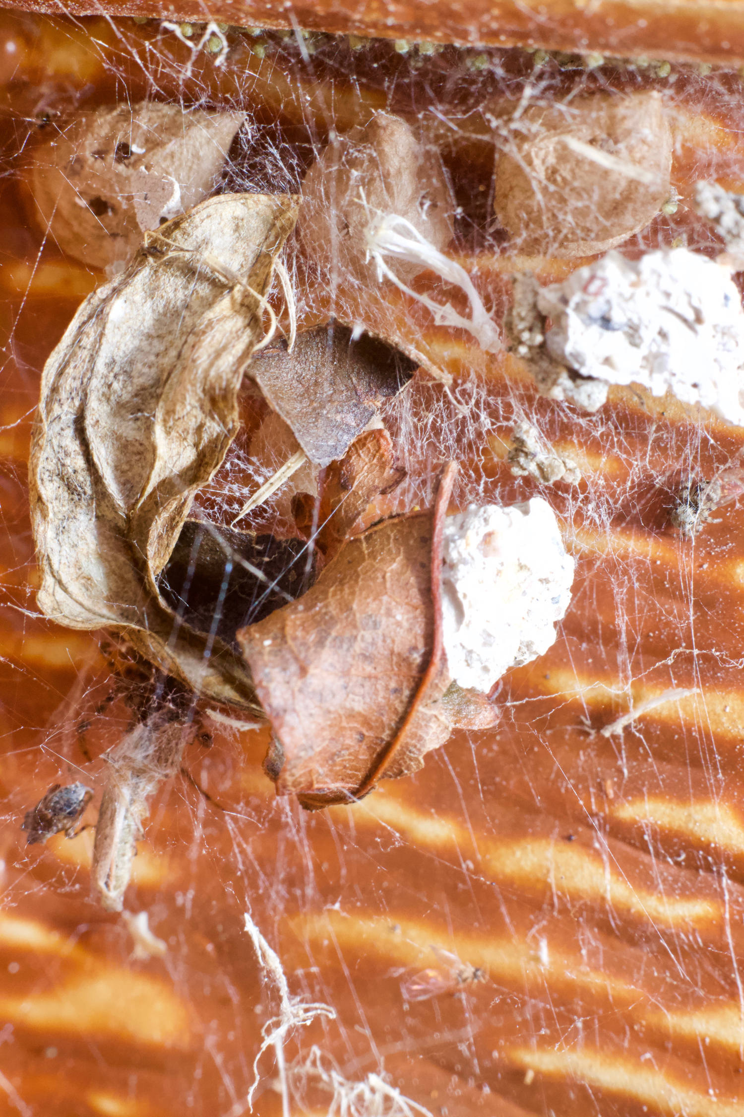
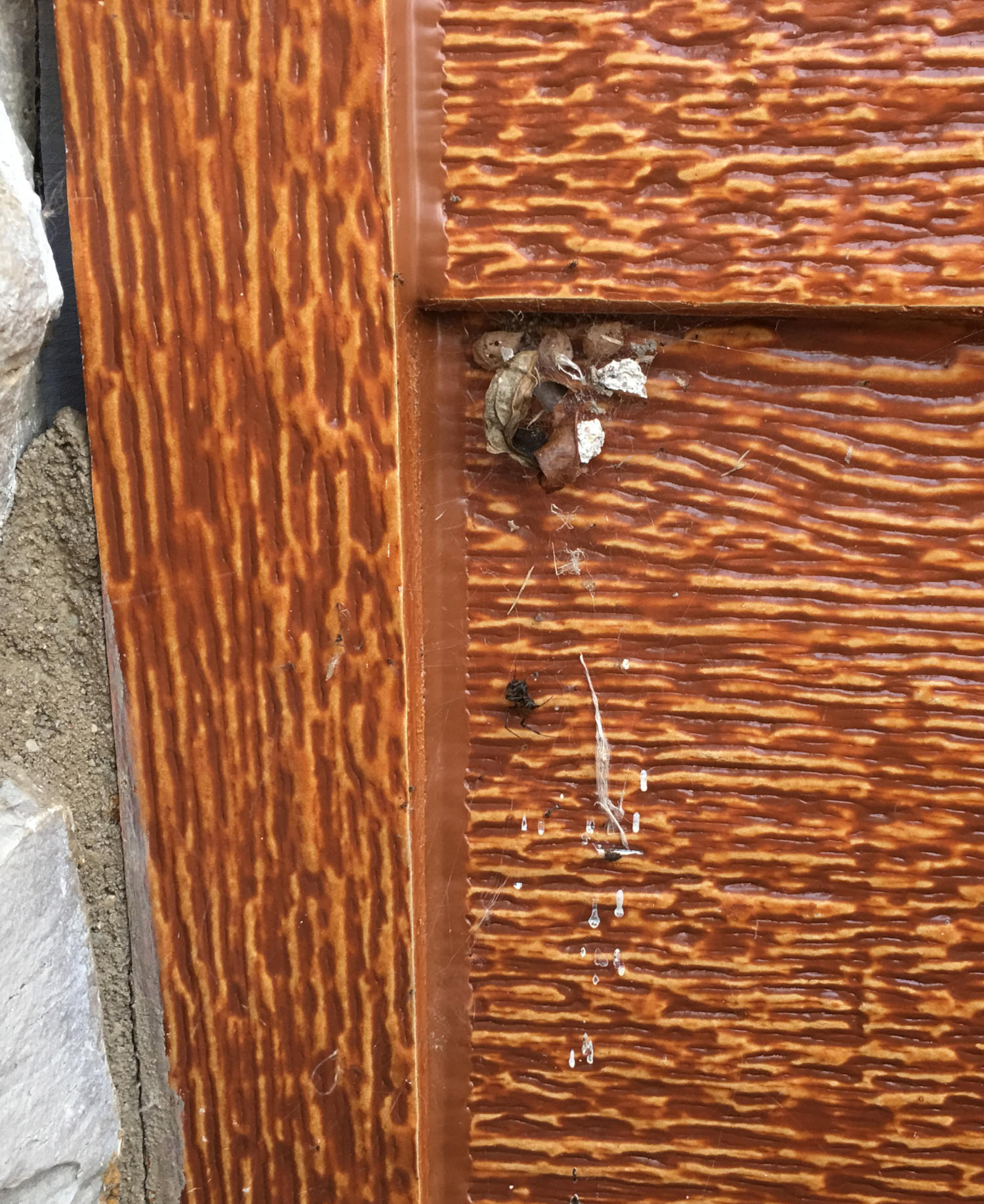
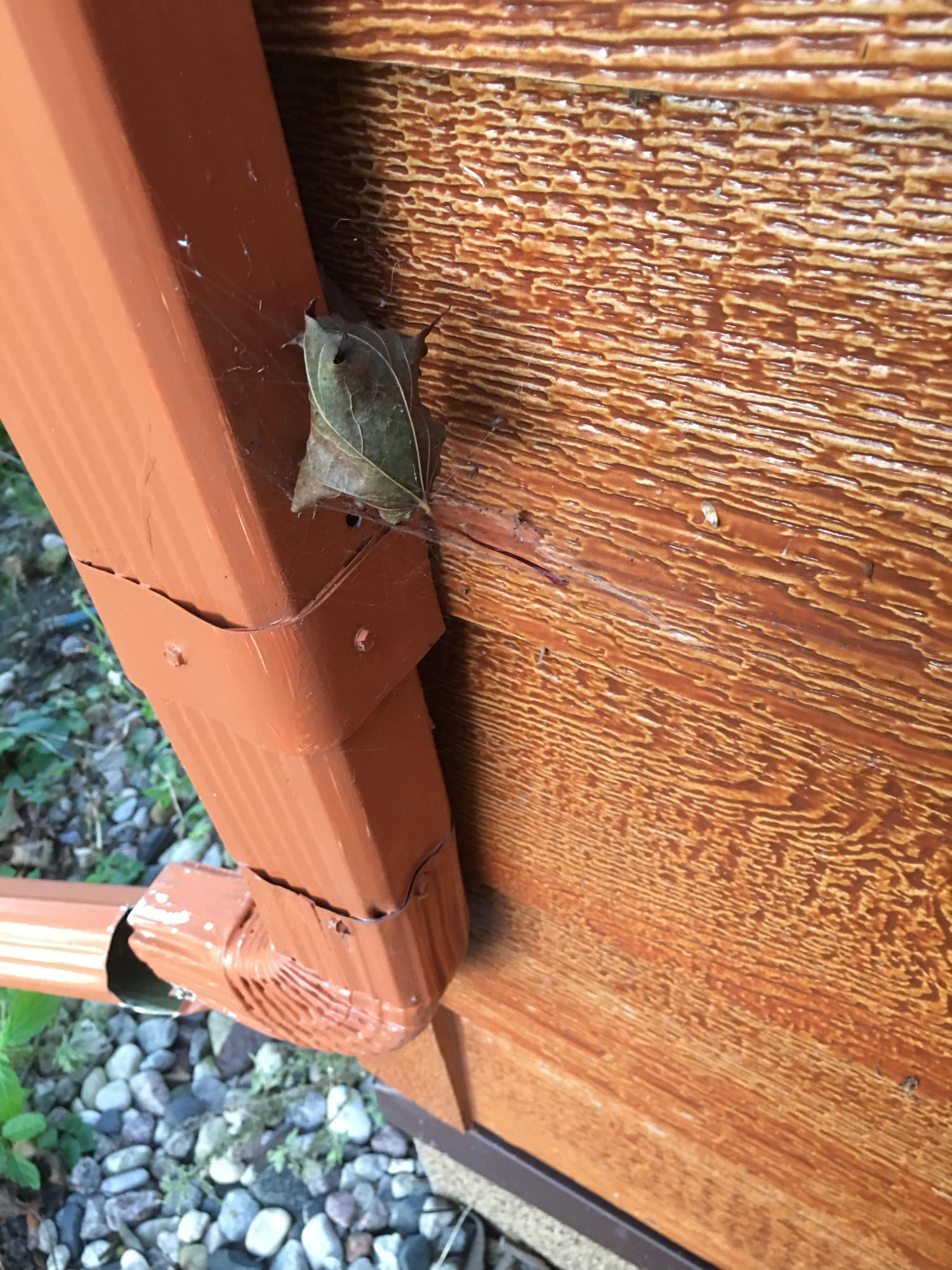
So I stopped by this morning (how could I not?), and the photo doesn’t do it justice. It is spider paradise. It’s a spider commune. There are all kinds of bugs living in the compost, and all over above them is a dense communal spider web, packed with spiders. I’d suspected it from the first picture, but I stuck my face down there and confirmed it — Steatoda borealis, the Northern Combfoot, which I’ve occasionally found while prowling about town, but this was the Mother Lode. I got a few closeups of one of their number in their web.