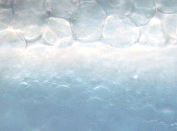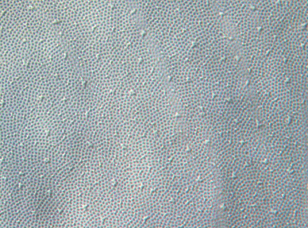
That strange tissue I showed in a previous post is…the chorion of the embryonic zebrafish. It’s homologous to a structure called the zona pellucida in mammals, and it’s also made of the same stuff: a collection of highly conserved glycoproteins called ZP (for zona pellucida proteins) that form a tight extracellular matrix around the egg. There are four groups of related proteins creatively called ZPA, ZPB, ZPC, and ZPX, and most are found in fish, frogs, birds, reptiles, mammals — so they really are universal.
One distinction is that only mammalian ZPs/chorions have the property of sperm recognition — in other groups the chorion acts explicitly as a barrier to sperm entry. Fish have a tiny funnel-shaped hole in their chorions called the micropyle at the animal pole, which is just big enough to allow a single sperm to enter, reducing the likelihood of polyspermy.
What’s also cool about the chorion is that it inflates and self-assembles. It lifts off the surface of the egg at fertilization and expands, and further, enzymes are released from cortical granules in the egg to harden and toughen the coat. Basically when the egg is fertilized it quickly blows up a fluid-filled bubble around itself.
In zebrafish, the chorion is thin and transparent, and relatively easy to tear and remove. Other fish species may differ; the first time I tried removing the chorion from medaka, it was like trying to rip through tough leather after after being used to peeling away soft toilet paper. Chorions may also be decorated with threads or spiky processes, especially in demersal (sinking) eggs that need to stick to rocks or grasses at the bottom of a stream. Zebrafish are rather mundane and plain in comparison.
There are complicated things going on in the chorion: it’s a barrier and a filter. It blocks some toxic or teratogenic agents — there are some substances, like steroid-like plant alkaloids (cyclopamine, jervine) that are much more potent if you remove or even just tear a small hole in the chorion.
So about that photo: you are looking at a very thin sheet of a glycoprotein matrix that forms a kind of eggshell around the embryo. Most of the time I just rip it off and throw it away, but in this case I was scanning embryos and left it on, and as always, it struck me as lovely and intricately patterned.
Bonsignorio D., et al., 1996. Structure and macromolecular composition of the zebrafish egg chorion. Zygote, 4(02), pp.101-108.
Iwamatsu T et al. 1995. Changes in chorion proteins induced by the exudate released from the egg cortex at the time of fertilization in the teleost, Oryzias latipes. Development, Growth & Differentiation, 37: 747–759.
Murata K et al. 2014. Identification of the Origin and Localization of Chorion (Egg Envelope) Proteins in an Ancient Fish, the White Sturgeon, Acipenser transmontanus. Biol Reprod 90(6): 132.
Rizzo E et al. Oocyte surface in four teleost fish species postspawning and fertilization. 1998. Braz. arch. biol. technol., Curitiba , 41(1):37-48.