What I taught today: induction in worms, early development in flies

Today was the due date for the take-home exam, which meant everything started a bit late — apparently there was a flurry of last-minute printing and so students straggled in. But we at last had a quorum and I threw worms and maggots at them.

The lab today involves starting some nematode cultures so I gave them a bit of background on that. They’re small, transparent hermaphrodites that can reproduce prolifically and will be squirming about on their plates this week. They’re models for the genetic control of cell lineage and also for inductive interactions: I gave them the specific example of the development of the vulva, in which a subset of cells in close proximity to a cell called the anchor cell develop into the primary fate of forming the walls of the vulva, cells slightly further away follow a secondary fate, forming supporting cells, and cells yet further away form the hypodermis or skin of the worm. I had them make suggestions for how we could test that the anchor cell was the source of an inductive signal, and yay, they were awake enough at 8am to propose some good simple experiments like ablation (should lead to failure of the vulva to form) or translocation (should induce a vulva in a different location). I also brought up genetic experiments to make mutants in the signal gene, in the receptors, and deeper cell transduction pathways.

All those experiments work in the predicted ways, and I was able to show them an epistasis map of the pathways. Two lessons I wanted to get across were that we can genetically dissect these pathways in model organisms, and that when we do so, we often find that toolkit Sean Carrol talks about exposed. For instance, in the signal transduction pathway for the worm vulva, there are some familiar friends in there — ras and raf, kinases that we’ll see again in cancers. And of course there are big differences: mutations in ras/raf in us can lead to cancer rather than eruptions of multiple worm vulvas all over our bodies, because genes downstream differ in their specific roles.

Then we started on a little basic fly embryology: the formation of a syncytial blastoderm, experiments with ligation and pole plasm manipulation in Euscelis that led to the recognition of likely gradients of morphogens that patterned the embryos. From there, we jumped to the studies of Nusslein-Volhard and Wieschaus that plucked out the genes involved in those interactions and allowed whole new levels of genetic manipulation. As the hour was wrapping up, I gave them an overview of the five early classes of patterning genes: the maternal genes that set up the polarity of the embryo; the gap genes that read the maternal gene gradient and are expressed in wide bands; the pair-rule genes that respond to boundaries in gap gene expression and form alternating stripes; the segment polarity genes that have domains of expression within each stripe; and the selector genes that then specify unique properties on spatial collections of segments.

And that’s what we’ll be discussing in more detail over the next few weeks.

Slides used in this talk

What I taught today: Nuffin’!

Nothing at all! I gave the students an exam instead! While I got a plane and left ice-bound Morris to fly to Fort Lauderdale, Florida! Bwahahahahahaha!

Sometimes it is so good to be the professor. And if ever you wonder why my students hate me with a seething hot anger, it’s because I’m such an evil bastard.

Here’s what they have to answer.

Developmental Biology Exam #1

This is a take-home exam. You are free and even encouraged to discuss these questions with your fellow students, but please write your answers independently — I want to hear your voice in your essays. Also note that you are UMM students, and so I have the highest expectations for the quality of your writing, and I will be grading you on grammar and spelling and clarity of expression as well as the content of your essays and your understanding of the concepts.

Answer two of the following three questions, 500-1000 words each. Do not retype the questions into your essay; if I can’t tell which one you’re answering from the story you’re telling, you’re doing it wrong. Include a word count in the top right corner of each of the two essays, and your name in the top left corner of each page. This assignment is due in class on Monday, and there will be a penalty for late submissions.

Question 1: We’ve discussed a few significant terms so far: preformation, mosaicism, regulation, epigenesis. Explain what they mean and how they differ from each other. Can we say that any one of those terms completely explains the phenomenon of development, or is even a “best” answer? Use specific examples to support your argument.

Question 2: Tell me about the lac repressor in E. coli and Pax6 in Drosophila. One of those is called a “master gene” — what does that mean? Is that a useful concept in developmental genetics, and is there anything unique to a gene in a multicellular animal vs. a single-celled bacterium that justifies applying a special concept to one but not the other?

Question 3: Every cell in your body (with a few exceptions) carries exactly the same genetic sequence, yet those cells express very diverse phenotypes, from neurons to nephrons. The easy question: explain some general mechanisms for how development does that. The hard part: answer it as you would to a smart twelve year old, so no jargon or technical terms allowed, but you must also avoid the peril of being condescending.

Wait…I’m going to have to fly back to Morris on Sunday, and then I’m going to have to read and grade all those essays! Aargh — they’re going to get their revenge!

What I taught yesterday: master genes and maps

On Wednesdays, I try to break away from the lecture format and prompt the students to talk about the science of development. We’re working our way through Sean Carroll’s Endless Forms Most Beautiful, and yesterday we talked about chapters 3 and 4.

Chapter 3 has an overview of basic molecular biology — transcription and translation, that sort of thing — and since these are junior and senior students who’ve already heard that a few times, we skipped right over it and they explained to me what master genes are, with specific examples of homeobox-containing genes like the Hox genes and Pax6. They caught on fast that what we call master genes are actually just transcription factors located high up in a regulatory hierarchy.

I think we also got across a less-than-naive idea of the evolution of Hox genes. There is a recognizable, conserved motif in each of these genes, but the proteins are far more than just their homeodomains, and can exhibit considerable variation — necessary functional variation, because the expression of different Hox genes are going to have distinct morphological consequences.

Chapter 4 has a general theme of maps and geography — what does it mean for a cell to be in a particular position and to have a particular fate? We also get into details. This is a very fly-centric chapter, and we get a picture of early development in the fly and the specific patterning and positional organization in the early embryo of that organism, with an introduction to many genes we’ll be hearing much more about during the course of the term. We also got enough information on vertebrate development that I could ask them to play the compare and contrast game: what’s different and what’s the same in fly and mouse development? I’m trying hard to be the Reese’s Peanut Butter Cup of development in this class: it’s so easy to say, “they’re the same!” and focus on common molecular mechanisms, or to say “they’re different!” and talk about the numerous quite radical innovations between them (especially in the fly, which is a weird, highly fine-tuned machine for rapid robust development). I’m trying to get across that both statements are absolutely true, and they really taste great together.

Friday is their first exam. Next Monday, class will be an overview of nematode development, to prime them for the lab exercises for the next two weeks which will be all about photomicrography of worm development and behavior, and also more details about early fly embryology to get them prepared for a couple of weeks of nothin’ but flies. I also warned them that next Wednesday we’ll be discussing chapter 5 in Carroll, just chapter 5, because I’ve found in the past that that’s usually the brain-clogger chapter, with all its talk of boolean logic and gates and circuits.

What I taught today: Axis specification

We began today with chocolate. Always a good thing at 8am, I think — so I brought a candy bar to class. Then I told the students that I loved and respected them all equally and that they all had equal potential, but that I was going to mark just one person as special by giving them that candy bar*. So I asked them how I could decide who should get it, telling them right off that dividing it wasn’t an allowed solution, and that yes, this could be an openly unfair process.

There were lots of suggestions: we could do it by random chance. I could throw it into the middle of the room and let them fight over it. We could analyze everyone’s DNA and give it to the most average person…or the most genetically unusual. I could just give it to the first person to raise their hand, or the person closest to me, or the person farthest from me. We could have a competition of some sort, and the winner gets it. I could give it to the person who wants it most, or who needs it most.

The point I was making is that this is a common developmental problem, that you have a potentially uniform set of cells and that somehow one or a few have to be distinguished as different, and carry out a different genetic program than another set of cells. One cop-out is to invoke mosaicism: that is, they aren’t uniform, but inherit different sets of cytoplasmic determinants that make them different from the very beginning, but that even in that case, these determinants aren’t detailed enough to specify every single cell fate in most organisms. Even with an initial prepattern, you’re eventually going to end up with a field of cells, like the dorsal side of the fly wing, and within that uniform field, some cells will have to be programmed to be epithelial, others to be bristles, others to be neurons. And that means that in every organism, even the most classically mosaic, you’ll reach a point where cells have to process information from their environment and regulate to build differential structures.

And with that I went on to talk about some animals that were judged as being mostly mosaic in character: molluscs, tunicates, echinoderms, and nematodes. Even here, these animals all required complex molecular interactions to build their embryos.

For example, I’d earlier used echinoderms as classic examples of regulative development. You can dissociate them at the 4-cell stage and each blastomere can go on to build a complete embryo. But at the 8-cell stage, when the cleavage plane separates an animal half from a vegetal half, that’s no longer true: the top four cells when isolated are animalized, forming only a ciliated ball, while the bottom four cells are vegetalized, only making a static blob with a bit of a skeleton inside. Clever experiments can quantitatively juggle these cells around, removing just the bottom 4 cells (the micromeres) at the 16-cell stage, or assembling composite embryos with different ratios of the different tiers of cells, and get different degrees of development. Even when you’re discussing an organism in which you’d call the pattern of development mosaic, it absolutely depends on ongoing cell:cell signaling at every step, and the final form is a consequence of interactions within the embryo. It’s a mosaic-regulative continuum.

I also described very superficially the work of Davidson and Cameron on specification events in echinoderms. These interactions can be drawn as a kind of genetic circuit diagram, where what you’re seeing is the pattern of genes being switched on and off. We can describe a cell type as the output of mappable gene circuitry, and we can even identify modules of networks of genes associated with a particular kind of cell, and that we can also see a limited number of genes that mediate interactions.

Next week I promised to start going into more detail, when we start talking about early fly development and axis decisions. The next class we’re actually going to switch gears a bit and discuss Sean Carroll’s Endless Forms Most Beautiful.

Slides used in this talk (pdf).

*Yeah, I lied again. I brought enough candy bars for everyone, and after we’d generated a list of ways to share just one, I gave them to everyone. They’ll never trust me again.

What I taught today: Position and Polarity

You really can’t teach a class by lecturing at them…especially not an 8am class. But sometimes there is just such a dense amount of information that I have to get across before the students know what to ask that I have to just tell them some answers. My compromise to deal with this eternal problem is to mix it up; some days are lecture days, others are discussion days. And today was a discussion day.

I’ve been talking at them for the past two weeks, basically working to bring them up to a 1950s understanding of the field of developmental biology, with a glimmering of the molecular answers to come and some of the general concepts, so that they’re equipped to start thinking about the contemporary literature. So I had them read this paper before class:

Kerszberg M, Wolpert L (2007) Specifying Positional Information in the Embryo: Looking Beyond Morphogens. Cell 130(2):205–209.

And then today they got into small groups and tried to explain it to each other. I primed them by suggesting that they try to define the terms positional information, gradient, morphogen, and polarity, and mentioned that I was playing a dirty trick on them, giving them a paper to introduce a basic concept that at the same time was pointing out some of the difficulties and problems of the idea, so I expected them to also do some critical thinking and question the concepts.

So they went at it. It went well; they had some lively conversations going on, which I always worry won’t happen with early morning classes. I find it helpful to ask students to try to poke holes in an idea, rather than just recite by rote what the paper says — it sends them hunting rather than gathering.

Several students noted that having a simple continuum of a molecule begs the question of how that gets translated into many discrete cell types; why does having one concentration of a morphogen make a cell differentiate into a thorax, while a very slightly lower concentration means it differentiates into an abdomen? It’s all well and good to suggest that a couple of overlapping gradients can specify position, like laying out a piece of graph paper with coordinates on it, but it doesn’t explain how that gets translated into position-specific tissues. I was most pleased that several of them, while groping for an answer, related it to the lac operon in E. coli, and brought up the idea of thresholds of gene activation. Yay! That so sets up future discussions about early fly embryogenesis, where that is exactly the answer.

I think they also got the idea that an explanation for general specification of body parts, for example, may not apply for explaining polarity within a body part; we may have to think about some kind of hierarchy of regulation, where we progressively partition the embryo into smaller and smaller units, with different mechanisms at different scales. They might be catching on to the depths of the problems to come.

Another way the paper primed the students was that it very briefly introduces a whole bunch of specific molecules: dpp, bicoid, sonic hedgehog, activin. They got a very general idea of the broad roles these molecules play, all part of my devious grand plan. When we start talking about the details of how animals set up dorsal-ventral polarity, for instance, and dpp/BMP start coming up in more specific contexts, I want them to be familiar old friends — molecules they already knew casually and informally, and now see doing very specific things, and interacting with another set of molecules, which now also joins their circle of pals. Before I’m done with them, they’re all going to regard these developmental signals and regulators as part of their family!

What I’m also going to teach today: a little image processing

My development class also has a lab. The last few weeks have all been teaching them how to get good optics on a research scope, and how to take photomicrographs with my PixeLink camera system. Today I’m going to show them how to appropriately process an image for publication, so they’ll learn a few digital enhancement tricks and a few ethical rules. I lay down a few laws about using image processing on scientific data:

What you must do to your image:

  • You must archive the original data and work with a copy. If I ask to see the original after you’ve enhanced the image up the wazoo, you better be able to show it.

  • You must document every step and every modification you make. You’re going to describe everything either on the image itself or in a figure legend; if this were to be published, you’d probably include it in the Methods section.

  • You must explain the scale and orientation of the image. The scale is usually shown by including a scale bar; orientation may be shown either by including annotations (text describing landmarks in the image) or an explanation in the figure legend, such as that it is a sagittal or horizontal section.

  • You must save the image in a lossless format, such as .png or .psd or .tiff. Do not save it in a lossy format like .jpeg, which can add compression artifacts.

What you may do to your image:

  • You may crop and rotate the image.

  • You may adjust the contrast and brightness for the whole image.

  • You may carry out simple enhancements, like applying a sharpening filter or unsharp masking, to the whole image…but remember, document everything!

  • You may splice multiple images together to produce a photomontage; you can also insert panels with enlarged or otherwise enhanced regions of the image, as long as it is absolutely clear what you’ve done.

What you may not do to your image:

  • You must not carry out selective modifications of portions of the image; you cannot sharpen the cell you care about and then reduce the contrast for other regions, for instance. You should not burn or dodge regions of the image.

  • No pixel operations or retouching: you are not allowed to go into the image and paint your data into existence!

We do a lot of this preliminary basic stuff because I run the course out of my research lab, rather than a student lab. I want to make sure they’re not going to break anything, and also that they know how to do good imaging, a skill they’ll find useful in other courses and in research (years ago when I taught this stuff, we’d also do black&white darkroom work — nobody does that any more, so now it’s all photoshop). The goal is to get them all able to churn out lovely photographic data, so later I can just hand them some nematodes or fruit fly embryos and tell them to do their own experiments and observations, just show me the pretty pictures when they’re done.

It’s a good life, being able to sit back and let students bring me gifts of biological beauty. I think they’ll also be posting some of these to their blogs.

What I taught today: gene regulation and signaling

Today was more context and a bit of a caution for my developmental biology course. I warned them that we’d be primarily talking about animals and plants (and mostly animals at that), but that actually, all of the general processes we’re describing are found in bacteria and other single-celled organisms — that in a lot of ways, microbiology is actually another developmental biology course. Yes, I went there: developmental biologists tend to be imperialists who see all the other sciences as mere subsets of the one true science. Of course, you could also take that as developmental biology being a synthetic discipline that steals bits and pieces from everywhere…

So the primary lessons today were reviews of stuff these students should have gotten in cell and molecular biology, with bits of biochemistry and microbiology thrown in. We talked about genes getting switched on and off, and the example I used was the classic: the lac operon in E. coli. What? You don’t know about it? It’s a beautiful system, in which the bacterium switches on the genes needed for digesting the sugar lactose only when the sugar is available in the environment. I showed this nice little 3 minute video:

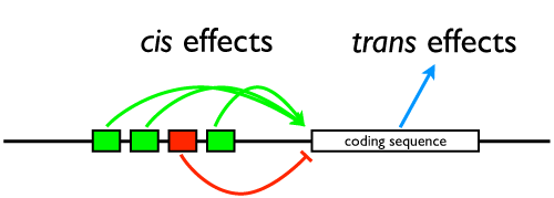
Switching genes on and off? That’s development! It gets a little more intricate in multicellular animals, but all of the fundamental logic is right there in E. coli: activators and repressors, positive and negative feedback, the boolean logic of gene regulation. I also mentioned that when we’re reading Carroll’s Endless Forms Most Beautiful, he’s going to make a big deal out of exactly this kind of regulation, but I want them to remember that it’s not unique to butterflies or fruit flies or frogs, it’s a common theme in all kinds of diverse cells.

We then talked about cell signaling. How does a cell know which genes are supposed to be off and on? It interacts with its environment (as in the lac operon) or its neighbors to make decisions about activity. To illustrate that, we went through quorum sensing and biofilms — again, cell signaling is not unique to animal development, it was all worked out in principle in single-celled organisms. I gave them a little foreshadowing and mentioned that we’d be discussing Sonic Hedgehog and Notch and Delta later in the course, classic examples of signaling in multicellular systems, but in bacteria we have things like hapR and AHL signaling.

Finally, I raised the issue of a phenomenon we’ll be talking about on Wednesday: patterning. Why aren’t your arms growing from your hips, why don’t you have fingers on your feet instead of toes, why are your eyes paired and on the front of your head? Because there is positional information in the embryo that can be read by cells and tissues and lead to development of appropriate structures in their proper places. But once more, this is not unique to multicellular animals. A paramecium, for instance, is not a generic blob, but has a definite shape and orientation; it has organelles in predictable places, and is covered with a nearly crystalline lattice of cilia with specific axes of orientation. I showed them choanoflagellates and pointed out that these protists, representing a multicellular precursor, had a specific shape and a collar organ in a specific functional location: how do they know how to do that?

That’s the question we’ll be asking next. I warned them too that I won’t be lecturing at them on Wednesday, so they’d better have their morning coffee. I’m expecting them to read a review paper on positional information in embryos (pdf), and I’m going to make them explain it all to me for a change.

Slides for this talk (pdf)

For Wednesday:

Kerszberg M, Wolpert L (2007) Specifying Positional Information in the Embryo: Looking Beyond Morphogens. Cell 130(2):205–209.

New bloggers for Science!

As is my custom, my upper level courses have an expectation that students will do this blogging thing. They’re just now getting set up so there isn’t a lot of content yet, but here’s the current list of student web pages. Cruise on by and talk to them!

What I taught today: a little old-school history of embryology

This is an abbreviated summary of my class lecture in developmental biology today. This was the first day of class, so part of the hour was spent on introducing ourselves and going over the syllabus, but then I gave a lightning fast overview of the history of developmental biology.

Classical embryology began with Aristotle, whose work was surprisingly good: he approached the problem of development with relatively few preconceptions and fairly accurately summarized what was going on in the development of the chick. Most of this old school embryology is descriptive and was really a narrow subset of anatomy, but there were a few major conceptual issues that concerned the old investigators, in particular the question of preformation (the plan of the embryo is laid out in the egg) vs. epigenesis (the plan of the embryo emerges progressively). Aristotle, by the way, was on the right side of this debate, favoring epigenesis.

In the 19th century, development was seen as a progressive process that paralleled the hierarchical organization of nature — that is, developmental biology, what there was of it, was coupled to the great ladder of being. This is not an evolutionary idea, but reflects the view that there was a coherent pattern of greater and lesser development that was part of a coherent divine plan for life on earth. The German ‘Natural Philosophers’ pursued this line of reasoning, often to degrees that now look ridiculous in hindsight. In contrast, there were developmental biologists like Karl Ernst von Baer who wanted nothing to do with a cosmic teleology but instead preferred to emphasize observation and data, and simple minimal hypotheses.

In the late 19th century, developmental biology split into two directions. One was a dead end; Ernst Haeckel basically lifted the explanatory framework of the natural philosophers, replaced divinity with evolution, and tried to present development as a parallel process to evolution. Von Baer had already demolished this approach, and despite a few decades of popularity Haeckelian recapitulation died as a credible framework for studying evolution in the early years of the 20th century. The other direction developmental biology took was Wilhelm Roux’s Entwicklungsmechanik, or experimental embryology. This was an approach that largely eschewed larger theoretical frameworks, and focused almost exclusively on observation and experimental manipulation of embryos. It was a successful discipline, but also divorced mainstream developmental biology from the evolutionary biology that was increasingly influential.

As examples of Entwicklungsmechanik, I discussed Roux’s own experiments in which he killed one cell in a two-cell embryo and saw partial embryos result, an observation that fit with a preformationist model, but more specifically a mosaic pattern of development, in which patterns of development were encoded into the cytoplasm or cortex of the egg. Those experiments were seriously flawed, however, because the dead cell was left attached to the embryo, and could have deleteriously affected development. The experiments of Hans Driesch were cleaner; he dissociated embryos at the four cell stage, cultured each blastomere independently, and discovered that each isolated cell developed fully into a complete, miniature larva.

Driesch, unfortunately, interpreted these results to imply that there was an entelechy, or guiding intelligence outside the embryo, and that the only conceivable explanation was the existence of purpose behind embryology. This was also a dead end; the modern explanation for the phenomena is that they regulated, that is, that cells determine their fate by interacting with one another, rather than some kind of cosmic plan. And that’s really going to be a major focus of this course: how do cells communicate with one another, how are genes regulated to set up coherent and consistent patterns of gene expression that produce the organized cell types we find in an adult multicellular plant or animal?

That set up the next lecture. Entwicklungsmechanik, while representing a solid and productive research program, quickly reached its limits, because what we really needed to examine were those patterns of gene expression rather than trying to infer them from observations of morphology. The big breakthrough was the melding of developmental biology and molecular biology — most of the modern developmental biology literature focuses on examining interactions between genes. So on Wednesday we’ll get another fast overview of the molecular genetics research program, and a bit of evo-devo.

Slide thumbnails (PDF)