
I could not leave the scarring of this lovely video unaddressed, so I made a video of my own.
Transcript below.
I was thrilled to run across this short time lapse video of some familiar neurons, cells that I became familiar with in the 1990s. Some ignorant Christian vandal has scrawled some words across it, but ignore those for now, and focus on just the beautiful developmental biology shown here.
There are some words that should be here, though. This video was appropriated from the Nikon Small World in Motion competition, where it won first prize in 2018. Here’s the announcement:
Nikon Instruments Inc. today unveiled the winners of the eighth annual Nikon Small World in Motion Photomicrography Competition. First place was awarded to Dr. Elizabeth Haynes and Jiaye “Henry” He for their video of a zebrafish embryo growing its elaborate sensory nervous system. The video reflects a time lapse of 16 hours and uses gentle light sheet technology to capture the whole zebrafish embryo in 3D, at a high temporal resolution. Dr. Haynes studies the role of kinesin light chain genes during the highly complex development of sensory neurons, while Mr. He specializes on developing microscopy technology to image living specimens at the best possible resolution.
Attribution is important, don’t you think? Now watch the show.
I recognized what was going on instantly. I had a graduate student, Beth Sipple, who did an analysis of the morphology and behavior of these cells for her doctoral work, so I spent a lot of time looking at videos and photographs of this sort of thing. I’ll take a moment to orient you.
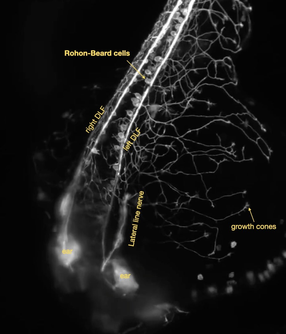
We’re looking at Rohon-Beard cells found in the dorsal spinal cord. These are cells that arise early in development, and the first thing they do is extend axons longitudinally in the spinal cord, pioneering the Dorsal Longitudinal Fascicle, or DLF, which is a major thoroughfare for sending sensory information to the hindbrain. This pathways is established by a daisy-chain of Rohon-Beard cells, so it forms very quickly and early in development. The cells then send a branch to the skin, with growth cones leading the way, where they form a kind of fishnet stocking of fibers that collect cutaneous sensory information.
One of the neat things about these particular cells is that they are thought to be an evolutionary holdover from the earliest chordate sensory system. They are kind of crude (but pretty): they are a diffuse information network that informs the animal that something has touched the left or the right side, with apparently little specificity otherwise. Imagine that you put up a fence around your property that rings an alarm if anything breaches it anywhere; that would be useful, but it’s not very specific about where the breach occurred. Perhaps more useful would be a series of sensors for each sector of the fence, that rings an alarm that tells you exactly where the contact occurred.
That’s coming, but it’s not shown in the video. If the timelapse extended for a couple of days, you might see those specific sensors emerging. They’re called dorsal root ganglia, and there’s a pair for each segment of the animal. So there’s a developmental sequence of first forming a broad general sensory network that can tell left from right, and then a specific localized sensory network that can tell exactly where on the body the animal was touched. This is also an evolutionary sequence, where the original primary sensory system is superceded by a much more precise secondary system, the dorsal root ganglia, which also contain multiple sensory modalities.
Also in the video you see the lateral line forming. This is a structure that first forms in the head, and then migrates caudally, dropping off neuromast organs as it goes. I found this to be a useful structure for timing development, because it grows like clockwork, and you can estimate the age of the embryo by how far down the body the lateral line has grown. I’m kind of rusty at staging embryos — I did most of my work with this system 30 years ago — but it looks the embryo at the start of the video is about 18 hours post-fertilization.
It’s beautiful stuff to see. It’s one reason developmental biology is so enthralling — you can see it all happening in the lab, over and over again, and it’s all an outcome of evolutionary history.
But this is also where the words the Christian vandal scribbled on the video are so jarring. Why are they talking about wombs? This is a zebrafish embryo. It’s taking place inside of an egg, no womb involved. And not only did they fail to acknowledge the authors of the work, they are needlessly vague about the organism. It’s a zebrafish, not a human, but apparently they want to mislead you into generalizing all the lovely neuronal choreography to human embryonic development. Surprise, though: humans don’t have Rohon-Beard cells. We skip straight ahead to developing a sensory system built around dorsal root ganglion cells.
Also, do I need to point out that people don’t have lateral lines? That’s an aquatic sensory system for detecting movement and compression waves in the fluid environment, we don’t have that.
If this were a human embryo, it would be a very boring video lacking all of the cells and fibers seen here!
But sure, Christians, steal the superficial details of the natural world around us and use your ignorance of what’s going on to invalidly justify your god nonsense. I have to tell you, though, that a scientific appreciation of the phenomena occurring here is far deeper and far more rewarding, and isn’t explained by a Bible verse. Try learning something, rather than trying to shoehorn Nature into your archaic theology.
See the original video unmarred by Christian graffiti here.

Leave a Reply
You must be logged in to post a comment.