I’ve said before that I’ve got a tear in the lateral meniscus of my right knee, and that I’m supposed to get that patched up with arthroscopic surgery in less than two weeks. But my right knee is the good one, that until last summer never gave me any problems! It’s my left knee that has been a lifelong troublemaker: I dislocated it while shoveling rocks when I was 13 (child labor is bad, trust me on this) and again when I was in high school playing basketball against the Kent-Meridian High School varsity football team (they didn’t understand that tackling wasn’t part of the official rules.) Both were treated by wrenching my kneecap back into place, and putting me in a hip-to-ankle cast for 3 months. Kids, don’t injure yourself while living in the middle ages.
As long as I was going in for surgery on the right knee, the doctor figured we should check out the left. I had an MRI this week, and just got the text summary, which looks like it’s mostly normal, but with some minor funny business that I can’t tell if it’s in the normal range, or if I ought to get it repaired now, before I retire. I understand all the words, but lack the context to know what to do about it.
EXAM:
MRI KNEE LT WITHOUT CONTRASTINDICATION:
Meniscal injury, knee,r/o meniscus injury,Internal derangement of left kneeTECHNIQUE:
Multiplanar multisequence knee MRI without contrastCOMPARISON:
Prior radiographsFINDINGS:
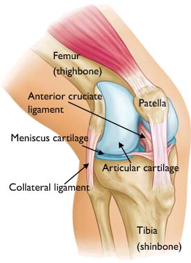
Bones:Patella and trochlear subchondral reactive edema with small cysts.
Normal marrow. Minor patellofemoral osteophytes.Ligaments, tendons:
ACL, PCL: Normal
Extensor mechanism: Proximal patellar tendinosis. Distal quadriceps normal.
Attenuated anterior fibers of MPFL suspicious for old proximal tear. Also
medial retinaculum. Minor thickening lateral retinaculum.MCL and post/med corner: Distended bursa versus ganglion cyst along the
posterior/medial corner between pes anserinus and semimembranosus.Lateral and post/lateral: Normal
Gastrocnemius tendons: Normal
Joint spaces:Small effusion. Minor reactive synovitis suprapatellar recess.
Diffuse patellar and trochlear cartilage loss mostly grade 2 and grade 3 with
small surface area grade 4 both sides. Small subchondral cysts.Low-grade chondrosis medial, lateral compartments
Soft tissues:
No Baker’s cyst. Diffuse grade 1 muscle fatty infiltration
Tibial, common peroneal nerves: Normal
Menisci:
Lateral:Free edge surface fraying midbody. No definite tear
Medial: Normal morphology, signal
Comment: Abnormal MRI findings very common in asymptomatic volunteers,
frequently not a source of symptoms. Many studies demonstrate meniscal tears
in up to greater than 50% asymptomatic volunteers, cartilage defects >24%, bone
marrow lesions up to 50%, 21% tendon abnormalities, prevalence increasing w
age. Nearly all pain-free adult knees have at least 1 MRI abnormal finding, so
MRI findings must be interpreted under supervision of expert clinical
assessment.Culvenor et al, Br J Sports Med 2019
Parkar & Adriaensen, Eur Radiol 2024
IMPRESSION:
1. Small effusion, reactive synovitis, patellofemoral cartilage loss
2. Mild patellar tendinosis
3. Suspected old partial tear MPFL retinaculum complex
4. Posterior/medial corner bursal distension versus ganglion or synovial cysts
That’s entertaining, and I appreciated that comment that “Nearly all pain-free adult knees have at least 1 MRI abnormal finding,” so I don’t feel any need to freak out. But I would raise my hand and say that I’m not pain-free, it’s been a chronic source of low-level pain for 50 years, and I don’t know what part of that is relevant to my situation.
I’ll talk to my doctor in the next few days to find out.

“Internal derangement” sounds bad.
You never know with medical terminology. I’m not going to get hung up on a few words here and there.
Most of those findings could be caused by the strain of favoring the knee due to injuring the other knee. Inflammation should dissipate when you get back to walking while distributing your weight evenly.
I would ask about this finding in particular-
Fraying would suggest that something hard is impinging on the menisci, and causing damage and pain.
Internal derangement is a catch all phrase the Orthopods use when something is wrong, the specifics elude them, but they need a diagnosis to order the test. Reminds me of the word idiopathic.
“Idiopathic” is a medical term used to describe a disease, condition, or symptom that arises spontaneously or has no known, identifiable cause.
The MRI report tells me the knee has been beat up (old). Multiple issues throughout but nothing crucial. Balding tires, still have tread but eventually???
“Knees” are one of the obvious clues that “if humans were DESIGNED, the designer is an IDIOT”.
Along with that whole “routing sewer output near recreation area” thing.
I feel your left knee pain. And the early 80s weren’t much better. I dislocated my patella when I was 10, but it popped back on its own. So treatment was a brace for 4 weeks and on my way.
From then until I was 17, when I had surgery, if I ran more than 100 meters or so it would give out again and I would find myself lying on the ground. Surgery solved the dislocations, but the damage was done. In recent years when I’ve had problems and its been looked at I get a conversation that goes something like: “here’s a knee MRI/x-ray/CT, see this area in the joint where the cartilage is? Now this is yours, you don’t have any left.”
MRI’s reports often make normal age-rleated changes sound terrifying.(especially MRI’s of the back). I’ve never seen a report say something like “Nearly all pain-free adult knees have at least 1 MRI abnormal finding,” before and that is actually really useful.information
Caveats: (1) investigations must be interpreted with a thorough understanding of the individual’s history and examination findings, (2) I am not an orthopaedic surgeon or a radiologist, and (3) I am now retired from clinical practice.
(1) “Internal derangement” is a catch-all term meaning there is some structural abnormality. It is not specific to any given injury or severity.
(2) I would think the best explanation of your left knee pain is osteoarthritis of the patellofemoral surfaces (i.e. the cartilage surfaces that allow the patella to slide over the knee as it bends). It fits the pattern of pain, the history of knee dislocations, and the MRI finding of “[d]iffuse patellar and trochlear cartilage loss mostly grade 2 and grade 3 with small surface area grade 4 both sides. Small subchondral cysts.” Even a small area of cartilage loss can cause a great deal of pain in an active joint.
(3) The purpose of the comment about meniscal tears being found in 50% of pain-free volunteers is to prevent over-diagnosis and subsequent unnecessary treatment. There is a long-standing problem of abnormal findings being blamed for chronic pain conditions when they may be completely unrelated. Similar studies exist for disc bulges in back pain. The point of putting this boilerplate paragraph in the report is to remind the reader that Abnormality X does not necessarily cause Symptom Y. Sometimes they don’t even correlate (I’ve seen back pain attributed to a disc bulge three levels too high to explain the symptoms). And it means invasive procedures should be carefully considered in the light of best clinical evidence.
You can find these sort of comments added automatically to many investigation reports, especially on tests that require careful interpretation (e.g. gene probes, coeliac serology) and a good understanding of sensitivity, specificity and predictive values.
dlpthomas@7–
Yes, a lot of medical terminology can be frightening (or just plain weird), often due to terms being used for a couple of centuries while language has moved on. “Degenerative change” on Xray just means wear-and-tear/osteoarthritis and is completely normal in older age groups, but it sounds awful, like MND or ALS/Gehrig’s Disease. There is movement to retire archaic terminology from medical communication, but I will miss pathology reports calling tissue injuries “insults”.
[OT]
chrislawson, medical terminology? It can surely amuse.
I had a MRI some time ago, got a ‘brain is unremarkable’ mention in the results.
(I took that as meaning not pathological or abnormal, but it’s not very flattering, is it? ;)
John Morales–
I had to train myself out of saying “they didn’t find anything on your brain scan.” Obviously not the message I was hoping to convey.
John Morales @10
Your unremarkable brain is unsurprising though I just remarked upon it 🤣
At least it wasn’t Abby Normal.
chrislawson @8
Thanks for the elaboration.
My own late teen injury involved a patellar subluxation if I recall correctly. Happened just that once under unusual circumstances…I was pushing a car in a gravel driveway and lost my step. One thing you don’t want to see is the orthopedist draw blood instead of clear fluid from your swollen knee. Been there.
Ironically my toy breed dog was susceptible to patellar subluxations. Vet recommended some preventative surgery, which I passed on. Kept her from getting too heavy instead. Vet admonished me when she had surpassed 9 pounds which I soon corrected. She would kick her back leg out and shake it once in a while if I walked her too much. I guess some larger breed have hip issues instead.
I’m around a decade behind PZ in age so I wonder if I’m getting a preview.
Get the bad one fixed first and get it back to good mobility, would be my advice. I’m not an orthopod, but I see no surgical indication for the left one. Maybe hyaluronic acid injections or somesuch.
I found that surgery made permanent the then-present state of pain and disability. I have two total knee replacements. I would give all of my current available cash to return to the previous state.