I’m getting a bit peeved at all this new technology. Why, back in the day when I was doing electron microscopy work, I’d spend days slicing up tiny fragments of zebrafish embedded in epon-araldite with an ultramicrotome, and I’d end up with hundreds of itty-bitty copper grids that I’d put in the EM one by one, pumping the chamber down to a good vacuum and scanning and focusing and taking pictures. On a film cartridge! That I had to take into a darkroom and process myself! Both ways, uphill, in the snow!
And now look at this. These guys can make a single thin section slice of the whole larva, throw it in a machine, and step back while it automatically scans everything, and then throws it all onto a digital image. Of course, in this case it took their machine 4½ days to shoot over 26,000 images and then stitch them all together, but that’s still far faster than I ever was. I’d slave away to get just one good picture of a chunk of synaptic neuropil maybe 20 micrometers on a side.
Damn. Now I know how John Henry felt.
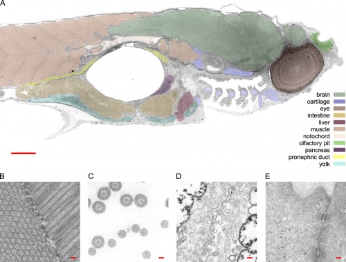
So here’s a transmission electron micrograph of a zebrafish at low resolution, just to help orient yourself. Ah, this is all familiar stuff; I spent most of my time hanging out in the nervous system, but those blocks of muscle (pink) on the left are always beautiful to look at. That big hole in the middle is the swim bladder, and the guts are slung underneath that. This is a parasaggital section — just off the midline — so it slices nicely through the eye (the big dark circle on the right) and also catches one nostril, above (that’s right, not where you might expect it*) and to the right of the eye. The scattered fragmentary stuff at the bottom, in front of the swim bladder, are sections through the pharyngeal arches. Take a look at the pretty cartilaginous rods sliced through in there.

The virtual slide was recorded at 120 kV with a magnification at the detector plane of 9460. A set of points was manually selected to outline the zebrafish and the convex hull of these points was used to define the data collection area. A total of 26,434 unbinned 4k × 4k images was collected with a FEI Eagle CCD camera (>8 s readout time full frame) in 4.5 d. The sample was maintained at −1 µm defocus throughout the whole data collection. The resulting slide of 1,461 × 604 µm2 consists of 921,600 × 380,928 pixels of 1.6 nm square each. The net data content of this slide is 281 Gpixel.
This is at high enough resolution that you can browse around the brain and find synapses and vesicles. Oh, you can’t see that? Go to the visual browser, and you’ll be able to zoom in and in and in. Easily. With no effort. Just glide on in there and find what you want.
Unlike my old experience with EM. Hey, if any of you have a time machine handy, could you grab one of these gadgets and drop it off for me in Eugene, Oregon, about 1982? Thanks.
*In case you’re wondering how nostrils can be above the eye, visualize a bulldog. Now grab it by the snout, and lift upward, stretching the face up so that a forward view is just a shot of the jaws with eyes on either side. Or, better than a bulldog, start with Admiral Ackbar.
Frank G.A. Faas, M. Cristina Avramut, Bernard M. van den Berg, A. Mieke Mommaas, Abraham J. Koster, Raimond B.G. Ravelli (2012) Virtual nanoscopy: Generation of ultra-large high resolution electron microscopy maps. Journal of Cell Biology 198:457-469 DOI: 10.1083/jcb.201201140.


Heh, know the feeling. I’m in the write-up stages of my PhD thesis, and my lab just installed a confocal microscope in the category 3 virus lab. After I spent more than a year faffing with tranfections and constructs to do live-cell imaging of my virus protein in action, we can now do it with full virus and I don’t have enough time left to try it (strict time limits on getting thesis in at my university, has to be in by the end of October).
Very cool tech tho, especially being able to splice together 26,000 images!
Still easier to be an IDiot. Just look at the fish, say reverently “It’s designed,” then “Acknowledge that it couldn’t possibly have evolved, infidel, or burn,” and write a book about it that many dunces will buy.
So see, ID is too superior, or at least a whole lot more money per hour for the right people, than all of this fiddly science junk!
Glen Davidson
Yeah, that’s very cool. Zoom in on those myomeres and check out the striations (sarcomeres)!
Glen D: Have you noticed that nobody seems to talk about ID anymore except (incessantly) for you? You seem to be stuck in 2005 or something. (Please respond with Tourettes-like vitriol.)
Are you new here? IDiots are savaged ’round here all the time.
Hell, Louisiana is currently considering handing over a good portion of their students to Intelligent Design nonsense through a private school voucher measure. We NEED to talk about it and fight it or we’ll be stuck with a new generation of unschooled citizens.
Re: nostrils above eyes.
There are some more familiar examples. Probably not necessary to torture a poor bulldog.
Dolphin: http://dolphin.lehocq.co.uk/MARTINGg.jpg
Brachiosaur: http://www.emc.maricopa.edu/faculty/farabee/biobk/brach10.jpg
To me, vampire bats kind of look like they might be heading in that direction:
http://images.nationalgeographic.com/wpf/media-live/photos/000/005/cache/common-vampire-bat_505_600x450.jpg
@ Disagreeable Me
But you’re ok with torturing Admiral Ackbar? You monster!
Don’t do that. It’s a trap!
You forgot to throw in a ‘now get off my lawn’….
If you find anyone with a time machine, I’ve got a few requests to add to your list.
Is it that thing about Heinrich Böllweiser? Everyone asks me to kill him, and I did, and then this Adolf Hitler guy who was even worse took over instead.
Tell me about it. I saved the Titanic from its infamous wrecking on The Needles, only for them to ram it into an iceberg.
What kind of preservative is used for this? Is it refrigerated? I’d imagine if the specimen was fresh and at room temperature that after four and half days it would be partially rotten.
That is so cool! Looks like I won’t be getting any work done this morning.
When I first read about the Atomic Force Microscope (and before I could get any technical details) it sure sounded suspicious to me – it didn’t involve all the hard work necessary for a Transmission Electron Microscope (the earlier Scanning Tunneling Microscope was another great invention that sounded a bit suspicious).
I know that problem. Cutting away for months on small larvae that are only 2mm in length, getting the slaps on small copper grids with one of your eyeleashes, staining them and then using a TEM to get the pictures on photo plates. My supervisor claimed that they still have better resolution than digital images though.
Ah, nevermind. A strong vacuum likely has excellent preservative qualities. Reading comprehension for the win.
Reminds me of Morris Louis with perhaps a bit of Helen Frankenthaler. The color part that is. Be nice printed really large on canvas. Or maybe fine line so you could get up close and see the synapses.
Heh. I was looking at submission instructions from a 1982 issue of a journal today.
For the love of Pete, use a clean typewriter ribbon!
Label your figures lightly with pencil on the backside!
Or better yet, submit only woodcuts.
Good to include John Henry by Belafonte. The complete 1959 recording where it is from is a classic.
@TomJ
Vacuum, nothing. I do not know exactly what they do for this extraordinary grade of microphotography, but for general cytology work a specimen is fixed with some pretty serious chemicals, set into wax and sliced very thin before being mounted on a slide. I can only imagine that for this it is vastly more complex and spoilage is the least of your concerns…
That purple stained organ to the bottom right of the swim bladder looks like testes not pancreas.
Oh boy, I am painting myself as a complete ignoramus here, but nostrils on a fish? I had no idea.
You damn kids get off my lawn!
I had to put a knife in that bastard Caligula, because PZ was too busy grinding lenses for the microscope.
@Gragara: Check out the nostrils on this beauty. Then, if you’re actually interested, you can start your ichthyological journey by reading up on fish anatomy on Wikipedia.
Thanks Lars. “The nostrils or nares of almost all fishes do not connect to the oral cavity, but are pits of varying shape and depth.” That’s what I thought, that they must be used for smell but obviously not for respiration. What I also learned is, that as (or before) air breathing evolved, nostrils developed a connection to the oral cavity, so as to enable the first air-breathing animals to breath while keeping most of their body under the surface.
Well, I’ve learned something new again. That’s always good. Also, I take it zebrafish are danio rerio ? I’ve always kept those in my aquarium, but they’re way too fast to see nostrils. Always on the move those little buggers are.